Time:2020-04-16 Reading:14556
First of all, do not
say whether these products contain real graphene, and do not say that there is
no data to support the various magical effects advertised by businesses, these
can at least more or less reflect the public's expectations of graphene, this
magical material.
In fact, in the
field of rigorous scientific research, scholars have been exploring more
possible applications of graphene. For example, can you imagine graphene being
used to 3D print artificial blood vessels? Recently, Alvaro Mata and other
researchers at Queen Mary University of London published a paper in the journal
Nature Communications, reporting a disordered protein and graphene oxide (GO)
co-assembly system, which can form capillary-like structures in a manner
similar to 3D bio-printing. Has some of the physiological properties of blood
vessel tissue, and is expected to be used in the lab to mimic key parts of
human tissues and organs.
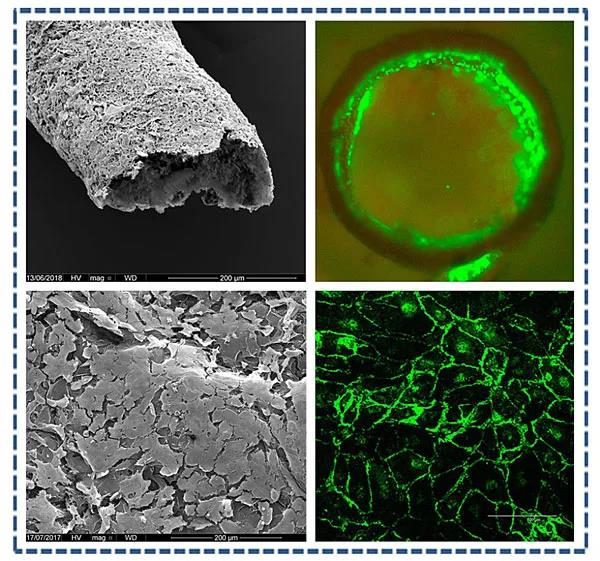
Self-assembly of a
vascular-like structure. Photo credit: Nat. Commun.
GO is rich in
oxygen-containing functional groups (hydroxyl, alkoxy, carbonyl and carboxyl)
and is often electronegative itself, so it can facilitate interactions with
different molecules -especially positively charged ones. The researchers'
choice of recombinamers to interact with graphene oxide is a disordered protein
-Elastin-like recombinamers (ELRs) -which is a class of recombinamers based on
the natural elastin motif Val-Pro-Gly-X-Gly (VPGXG, Where X can beany amino
acid other than proline), resulting in elastin-like peptides. Previous studies
have shown that these polypeptide molecules take on different molecular
conformations with temperature changes, can exhibit reversible phase
transitions, and have been used to make biocompatible materials. The three ELRs
involved in this paper are shown in Figure a below.
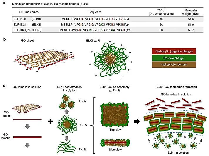
Molecular
construction and co-assembly principles. Photo credit: Nat. Commun.
Interestingly, when
the ELK1 solution drops into the GO solution, a multilayer film with a
thickness of ~ 50μm can be formed at the
interface around the droplet. The film is self-assembled by the charge
interaction of ELK1 and GO. Then, as the syringe moves while injecting the ELK1
solution, it can form a tubular structure, whose inner diameter is determined
by the size of the injection needle. The system can form capillary-like
structures through collaborative self-assembly in a culture medium where cells
are present, and the cells can be embedded in and on the wall of the tube.
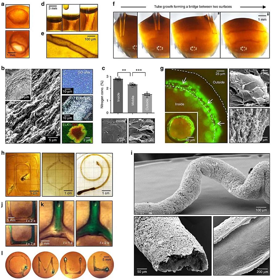
Self-assembly, structure
and properties of ELK1-GO. Photo credit: Nat. Commun.
If the ELK1 solution
is put into the extrusion 3D printer as "ink", you can freely
"print" different shapes of tubular structures in the GO solution. As
a result, the researchers printed tubes with different inner diameters,
different sizes, different torsion angles and different forks, which can
withstand a water flow of up to 12.5 mL/min for at least 24 hours. The highest
flow rate will produce a shear stress of 0.26 N/m2, which is not far from the
average shear stress generated by the common carotid artery (0.7 N/m2).
Since the reason for
the self-assembly comes from the strong interaction between ELK1 and GO at the
molecular scale, the solution concentration of both will certainly affect the
properties of the resulting tubular structure. The study found that the
interaction was strongest when the concentration ratio of the two was between
15 and 40, so during the "printing" process, the concentration of
ELK1 was maintained at 2%, while the concentration of GO was selected to be
0.05%, 0.10% and 0.15%. As expected, the strength, fracture strain and
toughness of the tubular structure all increased with the increase of GO
concentration. The tubular structures prepared with 0.10% GO solution had the highest
modulus of elasticity.
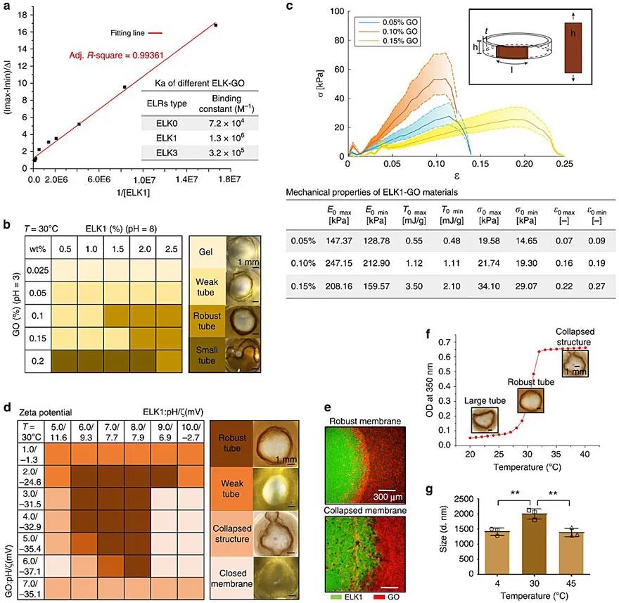
Molecular
interaction and mechanical properties test of ELK1 and GO. Photo credit: Nat. Commun.
At different
temperatures, the structural properties formed by the co-assembly of ELK1 and
GO are also different. It was found that the tubular structure obtained at the
transition temperature (Tt) of ELK1 at 30 °C
was more stable and clearer than that obtained at 4 °C
and 45 °C,
indicating a stronger interaction between ELK1 and GO at 30 °C.
The 3D conformation of ELK1 at different temperatures plays a key role in its
interaction with the GO sheet, which in turn influences the diffusion-reaction
mechanism that determines the performance of the resulting ELK1-Go tubular
structure.
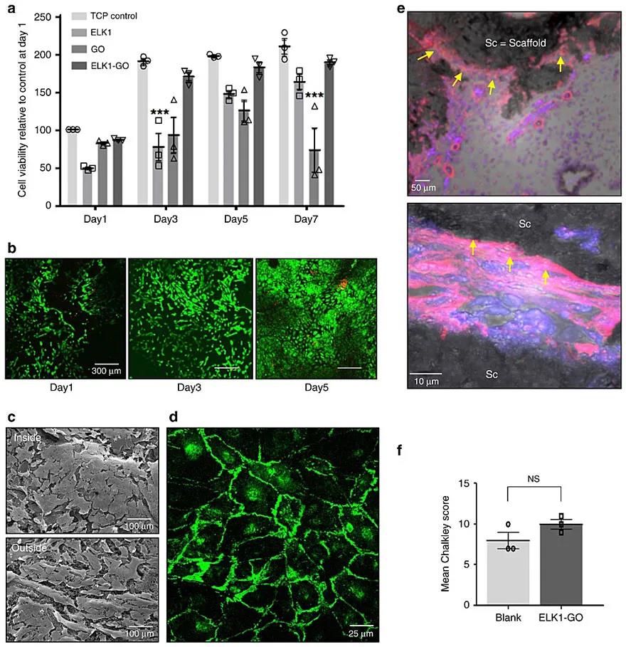
The cytotoxicity and
biocompatibility of ELK1-GO were verified in vitro. Photo credit: Nat. Commun.
"People are
very interested in developing materials and manufacturing processes that mimic
nature. However, the ability to build functional materials and devices through
self-assembly of molecules has been limited ", Yuanhao Wu says [1],
"This study introduces a new method to combine proteins with graphene
oxide through self-assembly, which can be easily combined with manufacturing
techniques to build biofluid devices, Enabling researchers to replicate key
parts of human tissues and organs in the lab."
References:
1. Biomaterial
discovery enables 3D printing of tissue-like vascular structures
https://www.nottingham.ac.uk/news/biomaterial-discovery-enables-3d-printing
Article converted
from X-MOL (x-mol.com)